HAP Chapter 4: Osseous system
Topic: Classification of Joints
Joint is a junction between two or more bones or cartilages. It is a device to permit movement. With the exception of the hyoid bone, every bone in the body is connected to or forms a joint. There are 230 joints in the body.
Functions of joint:
- Hold the skeletal bones together.
- Allow the skeleton some flexibility so gross movement can occur.
- Make bone growth possible.
Classification of joints: Joint are classified into structural and functional.
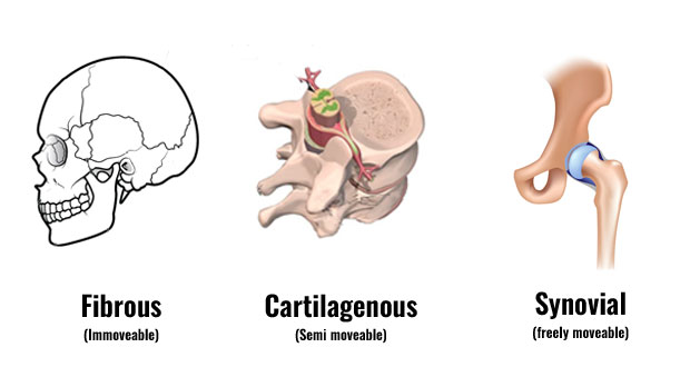
Structural classification is determined by how the bones connect to each other, while functional classification is determined by the degree of movement between the articulating bones.
Structural Classification of Joints:
A. Fibrous (Fixed):
1. Sutures a. Plane b. Squamous c. Serrate d. Dentate e. Schindylesis
2. Gomphosis
3. Syndesmosis
B. Cartilaginous (Slightly movable):
1. Primary Cartilaginous joints (Synchondrosis)
2. Secondary Cartilaginous joints (Symphysis)
C. Synovial Freely (movable):
1. Plane
2. Hinge
3. Pivot
4. Bicondylar
5. Ellipsoid
6. Saddle
7. Ball and socket

Fibrous Joints: Bones are joined by fibrous tissue/dense connective tissue, consisting mainly of collagen. The fibrous joints are further divided into three types:
1. Sutures or synostoses: Found between bones of the skull. In fetal skulls the sutures are wide to allow slight movement during birth. They later become rigid (synarthrodial).
Types of Sutures. (lambdoid suture)
2. Syndesmoses are joints where two adjacent bones are join together by a greater amount of connective tissue than in sutures in the form of interosseous ligaments and membranes. Eg-interosseous radioulanr joint, interosseous tibiofibular joint.
3. Gomphoses: It is a specialized fibrous joint restricted to fixation of teeth in alveolar sockets of the maxilla or mandible. The root of tooth is attached to the socket with in alveolus by periodontal ligament.
CARTILAGINOUS JOINTS: In this type of joint the bones are joined by cartilage. There are two types of cartilaginous joints:
1. Primary cartilaginous joints
2. Secondary cartilaginous joints
1. Primary cartilaginous joints (Known as “synchondroses”): Bones forming joints are connected by a plate of hyaline cartilage. These joints are immovable and mostly temporary in nature. This cartilage may ossify with age. Examples in humans are the joint between the first rib and the manubrium of the sternum, Joint between epiphysis and diaphysis of growing long bone.
2. Secondary cartilaginous joints (Known as “symphysis”): In these joints the articular surfaces of bone forming the joints are covered by thin plates of hyaline cartilage, which are connected by a disc of fibrocartilage. Eg. symphysis pubis, Intervertebral disc, Manubriosternal joint, Symphysis menti.
SYNOVIAL JOINTS: These joints possess a cavity and the articular ends of bones forming the joint are enclosed in a fibrous capsule. as a result, they are separated by a narrow cavity, the articular cavity, which is filled with a fluid called synovial fluid.
Characteristic features: The articular surfaces are covered by a thin plate of hyaline cartilage.
The joint cavity is enveloped by an articular capsule which consists of outer fibrous capsule and inner synovial membrane.
The cavity of joint is lined everywhere by synovial membrane except over articular cartilages.
The cavity is filled with synovial fluid secreted by synovial membrane which provides nutrition to articular cartilage and lubrication of articular surfaces.
Some joint cavity completely or incompletely divided by articular disc/ menisc.
Types of synovial joints: They are classified according to the types of movement possible or shape of the part of the bones involved.

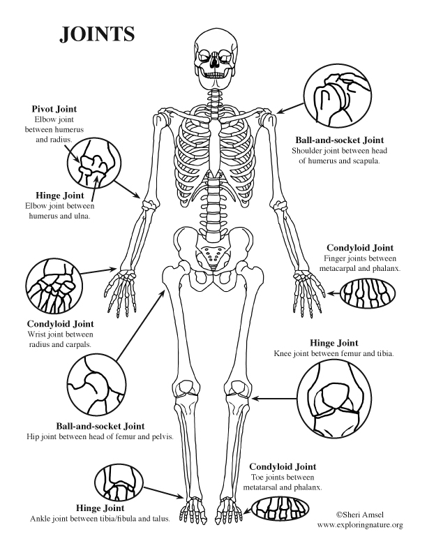
1. Ball & socket joint: The head of one bone is ball shaped which fits into cup shaped socket of another bone. This allows range of movement. E.g. shoulder joint, hip joint.
2. Hinge joint: The articulating ends form an arrangement similar to hinge on the door. The movement is restricted. e.g. elbow joint, knee joint, ankle joint.
3. Gliding joint: The articulating surfaces glide over each bet carpals (inter carpel bones), tarsal bones (inter tarsal bones).
4. Pivot joint: this joint allows the joint to rotate. e.g. the joint formed by axis & atlas allows the head to rotate & proximal & distal radio ulnar joint.
5. Condyloid joint: A condyle is a smooth projection of bone which fits on the depression of another bone. e.g. joint between Mandible & temporal bone, joint bet metatarsal & phalanges & joint between metacarpal & phalanges.
6. Saddle joint: The bones fit like man sitting on a saddle. e.g. the joint between first metacarpal and trapezium of wrist.