Chapter 4: Osseous system
Introduction:
- The human skeleton is the internal framework of the human body. It is composed of around 270 bones at birth – these total decreases to around 206 bones by adulthood after some bones get fused together. The bone mass in the skeleton makes up about 14% of the total body weight.
- The skeleton is the bony framework of the body.
- It forms the cavities and fossae (Shallow depressions or hollow parts) that protect some important body structures, forms the joints and gives attachment to the skeletal muscles.
Functions of Bones:
- Protection of soft organs.
- Support of the body (framework)
- Storage of minerals and fats (calcium)
- Blood cell formation.
- Provide the framework of the body.
- Give attachment to muscles and tendons.
- Permit movement of the body as a whole and of parts of the body, by forming joints that are moved by muscles.
- Form the boundaries of the cranial, thoracic and pelvic cavities, protecting the organs they contain.

Divisions of Skeletal System:
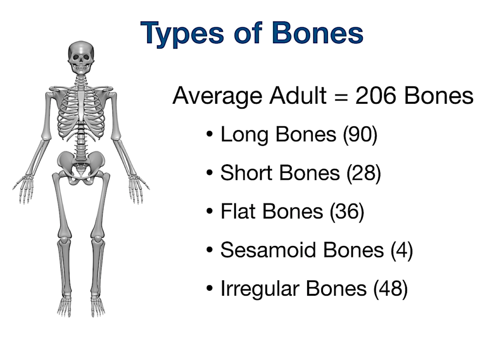
- Skeletal system contains 206 named bones which are divided into two main divisions as follows,
- The Axial Skeleton (80 bones)
- The Appendicular Skeleton (126 bones)
- The Axial Skeleton:
- Skull.
- Vertebral column.
- Sternum or breastbone.
- Ribs.
- The Appendicular Skeleton:
- The upper limbs and the shoulder girdles.
- The lower limbs and the innominate bones of the pelvis.

Bone and Types of Bones:
- Bone is a strong and durable connective tissue.
- It is made up of water, osteoid, bone cells constituting 50% part and remaining 50% of minerals like Calcium phosphate and Magnesium phosphate.
- Bones are classified into 5 types as,
- Long Bones.
- Short Bones.
- Irregular Bones.
- Flat Bones.
- Sesamoid Bones.

- Long Bones:
- These consist of a shaft and two extremities.
- As the name indicates the length is much greater than the width.
- Supports weight and facilitates movements.
- e.g. femur, tibia and fibula.
- Short Bones:
- Length is almost the same as width.
- Located in the wrist and ankle joints, these bones provide stability and some movement.
- e.g. Carpals in the wrist, tarsals in the ankles.
- Irregular Bones:
- As the name indicates they vary in shape and structure.
- They have a complex shape and protect the internal organs.
- e.g. the vertebrae.
- Flat Bones:
- They are flattened bones and also called “Sutural Bones”..
- Provide protection like shield and a vast area for muscle attachment.
- e.g. Skull bones, Sternum, Ribs, Pelvis etc.
- Sesamoid Bones:
- These are bones embedded in tendons.
- These small, round bones are commonly found in the tendons of the hands, knees, and feet.
- Their function is to protect tendons from stress and wear.
- e.g. patella (kneecap).
Bone cells:
- The cells responsible for bone formation are osteoblasts (these later mature into osteocytes).
- Osteoblasts:
- These are the bone-forming cells that secrete collagen and other constituents of bone tissue.
- They are present:
- in the deeper layers of periosteum.
- in the centres of ossification of immature bone.
- at the ends of the cartilages of long bones.
- at the site of a fracture.
- Osteocytes:
- As bone develops, osteoblasts become trapped and remain isolated in lacunae.
- They stop forming new bone at this stage and are called osteocytes.
- Osteoclasts:
- Their function is resorption of bone to maintain the optimum shape.
- This takes place at bone surfaces:
- under the periosteum, to maintain the shape of bones during growth and to remove excess callus formed during healing of fractures.
- round the walls of the medullary canal during growth and to canalise callus during healing.
- A fine balance of osteoblast and osteoclast activity is necessary for normal bone structure and functions.